Imaging Scans for Mesothelioma
Imaging scans are an important initial step in diagnosing mesothelioma. X-rays, CT scans and MRIs can help doctors identify mesothelioma tumors and the extent of disease. With other tests, imaging scans can help doctors diagnose mesothelioma and determine treatment options.
Learn About Mesothelioma Diagnosis in Our Free Guide Free 2026 Mesothelioma Guide
How Are Imaging Scans Used to Help Diagnose Mesothelioma?
Imaging scans are often the first step in the process of diagnosing mesothelioma, a type of cancer caused by asbestos exposure. Doctors may use a variety of imaging scans to help diagnose mesothelioma.
For example, a doctor may initially order a chest X-ray to look for any lung or chest abnormalities. The mesothelioma doctor may use a Positron Emission Tomography (PET) scan later in the process to help determine the patient’s stage of mesothelioma.
Depending on the type of scan, doctors can get information to:
- Estimate how the treatment is affecting the tumors
- Help understand how much cancer there is or how far it has spread
- Monitor a patient for cancer recurrence
- Show possible suspicious tissues or areas that might be cancer
Doctors may recommend several different imaging scans. While each has advantages and limitations, imaging scans alone do not provide enough information to allow a doctor to diagnose mesothelioma. Instead, doctors may recommend a blood test or biopsy. Testing a biopsy sample is the only definitive way to diagnose mesothelioma.
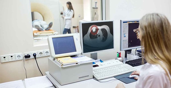
Radiology is a medical field that uses technology to get images of the inside of the body to help diagnose an injury or disease. Specialized doctors, called radiologists, use these images to help make a diagnosis. For example, radiology at a mesothelioma cancer center uses imaging tests to detect possible tumors in the thin lining around organs.


X-Rays: An Early Step in Mesothelioma Diagnosis
A doctor may order an X-ray early in the diagnostic process for mesothelioma. An X-ray sends electromagnetic radiation through a person’s body to capture images of structures inside the body.
X-rays are common medical imaging tests. Doctors often use X-rays as first-level diagnostic tools to see inside the patient’s body and discover any abnormalities.
X-rays usually take no longer than 5 – 10 minutes. However, some specific X-ray procedures, such as those involving contrast dye, may take longer.
Doctors use X-rays to help rule out other illnesses. For example, patients with difficulty breathing or chest pain may have a chest X-ray. The chest X-ray can help narrow down the cause of the patient’s symptoms from several conditions, including pneumonia, emphysema and heart issues.
- Standard X-rays take 5 – 10 minutes to complete, but the results may take time to review.
- X-rays use low levels of radiation, so they are unlikely to cause serious side effects.
- The radiation from one chest X-ray amounts to about what people naturally get over 10 days of being on Earth.
- There are different types of X-rays, including some that use special contrast dyes to highlight a specific part of the body.
- Newer technology, called digital radiography, does not use film. Instead, it takes an X-ray image like a digital camera and saves it on a computer.
Can You See Mesothelioma on an X-Ray?
An X-ray can help a doctor detect abnormalities in many parts of the body. Those abnormalities appear white on the X-ray, and healthy tissues appear black.
For instance, a chest X-ray can help a doctor detect tumors or excessive fluid buildup around the lungs. This excess fluid is called pleural effusion, a common symptom of pleural mesothelioma.
While a doctor can see the abnormalities, they will not be able to diagnose them as mesothelioma. The only way to diagnose mesothelioma is by testing a biopsy sample.
Chest X-Rays (CXR)
In cases of pleural mesothelioma, chest X-rays are the most common type of X-ray. Doctors order chest X-rays to look for abnormalities in the chest and around the lungs. Some common things a doctor looks for in a mesothelioma chest X-ray include:
- Pleural effusion or pleural thickening
- Loss of space in the chest cavity
- Lung compression
These findings may suggest additional tests are needed. The patient’s doctor can explain more about chest X-rays for pleural mesothelioma.
Computed Tomography (CT) Scans
A CT scan, sometimes called a CAT scan, can also help doctors diagnose mesothelioma. Like X-rays, CT scans are commonly used to view organs, tissues and tumors. Instead of a single image, CT scans provide a series of images from different angles.
CT scans use multiple X-rays that doctors can look at individually or stack on top of each other to create a 3D model. Complete results from a CT scan can take 24 hours. Doctors may use a CT scan for patients with signs of pericardial mesothelioma.
Contrast material, or contrast dye, is a substance that helps show abnormalities in the body. Contrast dyes are given by injection or orally. Doctors may use them during CT scans, MRIs or X-rays.
Some CT scans use a dye, or contrast material, to enhance the captured images. The contrast material makes it easier for doctors to visualize specific organs or tissues.
CT scans provide clear, specific information on the imaged tissues. They are also relatively fast and capable of imaging small or large areas within a single session. As such, CT scans are useful in determining the stage of mesothelioma.
- Some mesothelioma specialists prefer the CT scan for staging pleural mesothelioma cases.
- A typical CT scan usually takes between 10 and 30 minutes, depending on if the scan needs a contrast dye.
- Results may take 24 hours or longer, as a radiologist has to review and analyze the image.
- Patients lie on a table that moves in and out of a ring-shaped scanner during the quiet imaging scan procedure.
- CT scans can help doctors determine if a mesothelioma patient is eligible for surgery.
- The radiation a person gets from a CT scan is about what they naturally get over 2 years of living on Earth.
What Does Mesothelioma Look Like on a CT Scan?
A CT scan of pleural mesothelioma may show tumors along the lining around the lung (the pleura). The CT scan may also show pleural thickening or effusion, which are both consistent with a pleural mesothelioma diagnosis.
X-rays and CT scans both provide images of structures inside the body. However, CT scans show greater detail and allow the doctor to see it in 3D. CT scans cannot diagnose mesothelioma, but they may help lead a doctor to that diagnosis.
For example, peritoneal mesothelioma CT scans may show tumors within the abdominal cavity lining. A CT scan may also reveal fluid accumulation within the abdomen (ascites) or tumors on the liver or colon.
After diagnosis, studying a CT scan can also help doctors determine the best mesothelioma treatment, such as whether surgery is needed or possible.
Magnetic Resonance Imaging (MRI) Scans
An MRI scan can help diagnose mesothelioma by creating detailed images of structures inside the body. An MRI may show organs, bones, blood vessels and soft tissues like muscle or tumors. Doctors use these images to evaluate whether further tests, such as a biopsy, are needed.
MRI scans use radio waves and a strong magnetic field. It’s a painless, non-invasive procedure that does not use radiation. Some MRI scans may use an injectable contrast dye to help make tissues appear more clearly in the images.
Depending on the area being scanned, an MRI can take anywhere from 15 minutes to an hour or longer. The doctor may be able to tell the patient how long they expect it to take based on the images needed.
An MRI scan effectively highlights abnormalities in the body, including tumors and cysts. MRI scans may also show doctors how far the cancer has spread and if surgery for removal is possible.
- Usually, patients do not have to prepare for an MRI, but the doctor will have complete instructions.
- An MRI machine is a large tube that holds a powerful magnet and does not use radiation.
- Patients uncomfortable with tight spaces can ask about an open MRI, which provides more space.
- Doctors use MRI scans to determine if the mesothelioma tumor has spread to the diaphragm or chest wall.
- An MRI scan will not diagnose mesothelioma but can help doctors determine if the cancer has spread after a biopsy confirms it.
Can MRIs Detect Mesothelioma?
While MRIs can help detect mesothelioma, they are not a primary imaging tool for diagnosis. MRIs provide detailed images of soft tissues and can identify how far the tumor has spread. Other imaging methods, like CT scans, are more commonly used to support an initial mesothelioma diagnosis.
Doctors have found MRIs to be helpful for planning surgery on pleural mesothelioma tumors. They also help doctors figure out the stage of the disease and manage treatment for malignant pleural mesothelioma.
Positron Emission Tomography (PET) Scans
PET scans show tissues and cells that use a lot of energy, like cancer cells. For this type of imaging scan, the patient receives radioactive sugar. Cells that use a lot of energy absorb more sugar than other cells. Thus, cancer cells appear brighter compared to surrounding tissues.
A PET scan cannot show if a tumor is mesothelioma, but it does show abnormal tissue. Doctors can use that information to help determine:
- The size, location and growth speed of the tumor
- Whether thickening found in a CT scan is likely to be from cancer or scar tissue
- If the cancer has spread, or metastasized, to other parts of the body, like lymph nodes
Doctors may also use PET scans for mesothelioma patients to measure the efficacy of treatment. For instance, doctors may use a PET scan after or during a patient’s course of chemotherapy to look for tumor progression.
- A PET scan finds cells that use sugar (energy) more quickly, such as cancer cells.
- Cancer cells show up as bright spots on PET scans.
- Doctors use PET scans to find cancer, see if and where it has spread and how well treatment is working.
- A mesothelioma PET scan may help a doctor determine if the thickening of the pleura or peritoneum is from scar tissue or tumors.
- Some PET scan machines can also do CT scans.
PET Scans vs. CT Scans
PET scans and CT scans are collected the same way, using a rotating cylinder that gathers pictures from all angles. These scans differ mainly in the information they provide.
- CT scans create still images of tissues and bones. PET scans show which tissues are using a lot of energy.
- PET scans use radioactive sugar, and CT scans do not.
- PET scans generally take longer than CT scans.
Ultrasounds
Ultrasounds do not use radiation like X-rays, CT scans or PET scans. Instead, they use sound waves to travel through a person’s body and bounce back when they hit organs. The sound waves bounce off structures at different rates, allowing the machine to create images.
Ultrasound waves cannot transmit properly through air, which is found in the lungs and abdominal organs. This means ultrasound produces inferior images of structures in the chest and abdomen.
However, a doctor might opt for an ultrasound in certain instances, such as:
- If a patient presents symptoms or shows abnormalities that suggest testicular mesothelioma
- If there is fluid buildup around the heart (this type of ultrasound is known as an echocardiogram), which may be consistent with pericardial mesothelioma
For mesothelioma, doctors may use an ultrasound to pinpoint where a tumor or excess fluid, like a pleural effusion, is in the body. They may also use ultrasound to help guide a needle during certain procedures, like a thoracentesis or biopsy.
- Ultrasounds are not as detailed as CT scans or MRI scans.
- Ultrasounds do not work well in tissues that hold air, like the lungs.
- Doctors often use ultrasounds to guide needles during procedures, like biopsies, to help collect samples.
- An ultrasound can show the direction and speed of blood flow, which can help identify tumors.
- Most ultrasounds do not require preparation, but patients should speak with their doctors for instructions.
- Most ultrasounds take between 20 and 30 minutes to complete.
Mesothelioma Image Testing by Type
Image testing varies by mesothelioma type. Certain imaging tests may work well for some mesothelioma types but not others. For example, doctors commonly use ultrasounds for testicular mesothelioma. However, they typically do not use them for peritoneal mesothelioma.
- X-ray: Chest X-rays and abdominal X-rays are both used.
- CT scan: Doctors may use a CT scan of the chest during diagnosis.
- MRI: Doctors may recommend an MRI for patients if they are being considered for surgical treatments.
- PET scan: Patients eligible for surgery may have a PET scan to help the doctors stage the cancer.
- Ultrasound: Doctors may use an ultrasound to find pleural effusions or to examine lymph nodes to determine if the mesothelioma has spread.
- X-ray: Testing includes abdominal X-rays, and may include chest X-rays.
- CT scan: Doctors may use a CT scan of the abdomen during diagnosis.
- MRI: Patients may get an MRI if the doctor thinks surgery and HIPEC, a type of multimodal treatment, would be beneficial.
- PET scan: While uncommon, a PET scan may be used to help stage the mesothelioma or monitor treatment.
- Ultrasound: Doctors do not generally use ultrasounds in peritoneal cases.
- X-ray: Doctors may order a chest X-ray but less often than CT scans or MRIs.
- CT scan: Doctors may use a CT scan of the chest during diagnosis.
- MRI: MRI scans are common among patients diagnosed with pericardial mesothelioma.
- PET scan: It is unclear if PET scans are used to diagnose pericardial mesothelioma.
- Ultrasound: Doctors use a type of ultrasound called an echocardiogram to find pericardial effusion or pericardial tumors. They may also use them to guide needles for pericardiocentesis and/or a needle biopsy.
- X-ray: Patients may have a chest X-ray to check for metastasis.
- CT scan: Because testicular mesothelioma is rare, not much evidence shows up on standard imaging scans. However, patients may have abdominal or pelvic CT scans.
- MRI: It is unclear if MRI is used to diagnose testicular mesothelioma.
- PET scan: It is unclear if PET scans are used to diagnose testicular mesothelioma.
- Ultrasound: Doctors use ultrasounds as one of the primary diagnostic tools for testicular mesothelioma. These ultrasounds may lead to additional procedures to definitively diagnose testicular mesothelioma.
What Are the Next Steps for a Mesothelioma Diagnosis?
Imaging scans can provide doctors with valuable information during and after a mesothelioma diagnosis. For instance, imaging scans may help doctors classify cancer spread or clinical stage using the TNM system. However, imaging alone is not enough to make a definitive diagnosis.
A mesothelioma doctor may use an imaging scan as one of the early tests during diagnosis. After doctors review the imaging scans, they may order other tests, including blood tests and a biopsy.
Testing tissue from a biopsy is the only definitive method of diagnosing mesothelioma. Once the doctor confirms the mesothelioma diagnosis, they will develop the patient’s treatment plan.
Imaging scans may be used again during treatment. These scans can help doctors monitor how well the treatment works, such as looking for any cancer progression after chemotherapy.
Common Questions About Mesothelioma Imaging
Does asbestos show up on a CT scan?
Do asbestos fibers show up on an X-ray?
What is the best imaging for mesothelioma?
Are image scans covered by insurance?
Sources
American Cancer Society. Tests for Mesothelioma.
American Cancer Society. Understanding Radiation Risk from Imaging Tests.
American Cancer Society. X-rays and Other Radiographic Tests for Cancer.
American College of Radiology. What Is a Radiologist?
Chandramohan A, Shah N, Thrower A, et al. Communicating imaging findings in peritoneal mesothelioma: the impact of “PAUSE” on surgical decision-making. Insights Imaging. 2021;12(1):174.
Chicago Consensus Working Group. The Chicago Consensus on peritoneal surface malignancies: Management of peritoneal mesothelioma. Cancer. 2020;126(11):2547-2552.
Godar M, Liu J, Zhang P, Xia Y, Yuan Q. Primary pericardial mesothelioma: a rare entity. Case Rep Oncol Med. 2013;2013:283601.
Johns Hopkins Medicine. Chest Ultrasound.
Johns Hopkins Medicine. Magnetic Resonance Imaging (MRI).
Katz SI, Straus CM, Roshkovan L, et al. Considerations for imaging of malignant pleural mesothelioma: a consensus statement from the international mesothelioma interest group. J Thorac Oncol. 2023;18(3):278-298.
Kim J, Bhagwandin S, Labow DM. Malignant peritoneal mesothelioma: a review. Ann Transl Med. 2017;5(11):236.
Mayo Clinic. Chest X-rays.
Mayo Clinic. CT scan.
Mayo Clinic. Mesothelioma.
Mayo Clinic. Positron emission tomography scan.
McGehee E, Gerber DE, Reisch J, Dowell JE. Treatment and outcomes of primary pericardial mesothelioma: a contemporary review of 103 published cases. Clin Lung Cancer. 2019;20(2):e152-e157.
MedlinePlus. Imaging and radiology.
National Cancer Institute. Computed Tomography (CT) Scans and Cancer.
National Cancer Institute. Malignant Mesothelioma Treatment (PDQ®)–Patient Version.
Nazemi A, Nassiri N, Pearce S, Daneshmand S. Testicular mesothelioma: an analysis of epidemiology, patient outcomes, and prognostic factors. Urology. 2019;126:140-144.
Volpi F, D’Amore CA, Colligiani L, et al. The use of chest magnetic resonance imaging in malignant pleural mesothelioma diagnosis. Diagnostics (Basel). 2022;12(3):750.
Free Mesothelioma Treatment Guide


Katy Moncivais, Ph.D., has more than 15 years of experience as a medical communicator. As the Medical Editor at Mesothelioma.com, she ensures our pages and posts present accurate, helpful information.

Annette Charlevois is a Patient Support Coordinator for Mesothelioma.com. For more than 20 years, she has helped thousands of mesothelioma patients and their families get the assistance they need.